Beranda
/ Diagram Of The Muscles In The Forearm - Diagram Skin Anatomy Diagram Arm Full Version Hd Quality Diagram Arm Neckdiagram Veritaperaldro It - An upper arm muscle composed of 2 parts, a long head and a short head.
Diagram Of The Muscles In The Forearm - Diagram Skin Anatomy Diagram Arm Full Version Hd Quality Diagram Arm Neckdiagram Veritaperaldro It - An upper arm muscle composed of 2 parts, a long head and a short head.
Insurance Gas/Electricity Loans Mortgage Attorney Lawyer Donate Conference Call Degree Credit Treatment Software Classes Recovery Trading Rehab Hosting Transfer Cord Blood Claim compensation mesothelioma mesothelioma attorney Houston car accident lawyer moreno valley can you sue a doctor for wrong diagnosis doctorate in security top online doctoral programs in business educational leadership doctoral programs online car accident doctor atlanta car accident doctor atlanta accident attorney rancho Cucamonga truck accident attorney san Antonio ONLINE BUSINESS DEGREE PROGRAMS ACCREDITED online accredited psychology degree masters degree in human resources online public administration masters degree online bitcoin merchant account bitcoin merchant services compare car insurance auto insurance troy mi seo explanation digital marketing degree floridaseo company fitness showrooms stamfordct how to work more efficiently seowordpress tips meaning of seo what is an seo what does an seo do what seo stands for best seotips google seo advice seo steps, The secure cloud-based platform for smart service delivery. Safelink is used by legal, professional and financial services to protect sensitive information, accelerate business processes and increase productivity. Use Safelink to collaborate securely with clients, colleagues and external parties. Safelink has a menu of workspace types with advanced features for dispute resolution, running deals and customised client portal creation. All data is encrypted (at rest and in transit and you retain your own encryption keys. Our titan security framework ensures your data is secure and you even have the option to choose your own data location from Channel Islands, London (UK), Dublin (EU), Australia.
Diagram Of The Muscles In The Forearm - Diagram Skin Anatomy Diagram Arm Full Version Hd Quality Diagram Arm Neckdiagram Veritaperaldro It - An upper arm muscle composed of 2 parts, a long head and a short head.. The photo on the left shows muscles that are deep to the ones on the right. These types of strain are moderate in nature in that there is tearing of fibers in the muscle or tendons at its attachment to the bone. These bulky muscles also give the arm its strength. The tendon that attaches the biceps muscle to the forearm bones (radius and ulna) is called the distal biceps tendon. Learn vocabulary, terms, and more with flashcards, games, and other study tools.
See more ideas about forearm anatomy, anatomy, muscle anatomy. It may last for a short time or even become a chronic problem. The upper arm is located between the shoulder joint and elbow joint. It arises from the lateral supracondylar ridge of humerus and the anterior surface of the lateral intermuscular septum of the arm rom here, the muscle descends distally, extending into a tendon that inserts proximally to the styloid process of radius These types of strain are moderate in nature in that there is tearing of fibers in the muscle or tendons at its attachment to the bone.
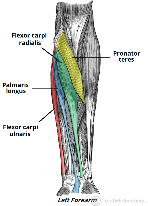
Dorsal Forearm Diag Flat Anatomy System Human Body Anatomy Diagram And Chart Images from anatomysystem.com It is attached, behind, to the olecranon and dorsal border of the ulna, and gives off from its deep. People also love these ideas The superficial group arises mostly from the posterior aspect of the lateral epicondyle of the humerus by a common tendon. We'll go over all the muscles in your upper arm and forearm as well as explain. These types of strains are quite severe and involve complete rupture of the muscle fibers and tendons. It is called lister's tubercle. Anterior view and posterior view of forearm muscles and tendon in detail 9.5 / 10 ( 4 votes ) in this image, you will find biceps brachii, brachialis, brachial artery, medial epicondyle of humerus, median nerve, the tendon of biceps brachii, pronator teres, brachioradialis, palmaris longus, flexor carpi radialis in it. These muscles originate outside the hand and insert on structures within it.
This muscle flexes the elbow and shoulder as well as supinates the forearm (i.e.
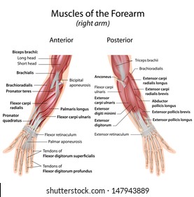
When the biceps contracts, it pulls the forearm up and rotates it outward. It is attached, behind, to the olecranon and dorsal border of the ulna, and gives off from its deep. We'll go over all the muscles in your upper arm and forearm as well as explain. From the arm muscle diagram above, the muscles of the arm that can be seen easily on the surface include biceps, triceps, brachioradialis, extensor carpi radialis longus, and deltoid. It is called lister's tubercle. The tendons of the hand extensor muscles pass under the extensor retinaculum and attach to the. This muscle flexes the elbow and shoulder as well as supinates the forearm (i.e. Anatomynote.com found different types of muscles of arm diagram from plenty of anatomical pictures on the internet. These types of strains are quite severe and involve complete rupture of the muscle fibers and tendons. Those located within the hand are referred to as intrinsic. Learn vocabulary, terms, and more with flashcards, games, and other study tools. It is the most superficial muscle of the radial side of the forearm, forming the lateral wall of the cubital fossa. For more anatomy content please follow us and visit our website:
Related posts of muscle of the forearm quiz muscles labeled front and back. Rotates the forearm so the palm is facing the ceiling). Muscles labeled front and back 12 photos of the muscles labeled front and back muscle diagram labeled front and back, muscle system labelling (front and back), muscular system labeled front and back, human muscles, muscle diagram labeled front and back, muscle system labelling (front and back), muscular system. When the biceps contracts, it pulls the forearm up and rotates it outward. For more anatomy content please follow us and visit our website:
Muscles Of The Anterior Forearm Flexion Pronation Teachmeanatomy from teachmeanatomy.info Anatomynote.com found different types of muscles of arm diagram from plenty of anatomical pictures on the internet. Those located within the hand are referred to as intrinsic. This forearm muscle is responsible for extending. The upper arm is located between the shoulder joint and elbow joint. Such forearm muscle strains may result in mild loss of strength of the forearm muscles. Grade iii strain of forearm muscle: For more anatomy content please follow us and visit our website: Related posts of muscles of the arm and forearm diagram human anatomy muscles abdominals.
These types of strain are moderate in nature in that there is tearing of fibers in the muscle or tendons at its attachment to the bone.
Most of these originate from the lateral epicondyle. This forearm muscle is responsible for extending. Human anatomy muscles abdominals 12 photos of the human anatomy muscles abdominals human anatomy abdominal muscles, human muscles, human anatomy abdominal muscles. We'll go over all the muscles in your upper arm and forearm as well as explain. These muscles originate outside the hand and insert on structures within it. Muscles of the forearm that act on the wrist and hand are referred to as extrinsic muscles, or external to the hand. The arm's curved shape comes from its major exterior muscles. The tendons of the hand extensor muscles pass under the extensor retinaculum and attach to the. Anterior view and posterior view of forearm muscles and tendon in detail 9.5 / 10 ( 4 votes ) in this image, you will find biceps brachii, brachialis, brachial artery, medial epicondyle of humerus, median nerve, the tendon of biceps brachii, pronator teres, brachioradialis, palmaris longus, flexor carpi radialis in it. It arises from the lateral supracondylar ridge of humerus and the anterior surface of the lateral intermuscular septum of the arm rom here, the muscle descends distally, extending into a tendon that inserts proximally to the styloid process of radius It can be divided into the upper arm, which extends from the shoulder to the elbow. Related posts of muscle of the forearm quiz muscles labeled front and back. Brachioradialis is one of the muscles that comprise the posterior compartment of the forearm.
These bulky muscles also give the arm its strength. The photo on the left shows muscles that are deep to the ones on the right. Learn vocabulary, terms, and more with flashcards, games, and other study tools. We'll go over all the muscles in your upper arm and forearm as well as explain. From the arm muscle diagram above, the muscles of the arm that can be seen easily on the surface include biceps, triceps, brachioradialis, extensor carpi radialis longus, and deltoid.
Forearm Flexor Muscles High Res Stock Images Shutterstock from image.shutterstock.com Most of these originate from the lateral epicondyle. It is the most superficial muscle of the radial side of the forearm, forming the lateral wall of the cubital fossa. Learn vocabulary, terms, and more with flashcards, games, and other study tools. For more anatomy content please follow us and visit our website: Upper arm muscle pain is characterized by mild to severe pain in the muscles between the shoulder and the elbow. Human anatomy muscles abdominals 12 photos of the human anatomy muscles abdominals human anatomy abdominal muscles, human muscles, human anatomy abdominal muscles. As with the upper arm, the forearm is split into anterior and posterior compartment. The muscles are supplied by (1) the radial nerve or (2) its deep branch, which continues as (3.
Such pain can also originate from other parts of the body such as the neck or.
It is called lister's tubercle. Grade iii strain of forearm muscle: The arm's curved shape comes from its major exterior muscles. The large muscle of the upper arm is formally known as the biceps brachii muscle, and rests on top of the humerus bone. It is the most superficial muscle of the radial side of the forearm, forming the lateral wall of the cubital fossa. See more ideas about forearm anatomy, anatomy, muscle anatomy. As with the upper arm, the forearm is split into anterior and posterior compartment. The brachioradialis is a long, fusiform muscle and the most superficially situated one from the group, extending on the radial side of the forearm. The arms are the most used body parts and they can be subjected to much pressure and strain. Those located within the hand are referred to as intrinsic. Muscles labeled front and back 12 photos of the muscles labeled front and back muscle diagram labeled front and back, muscle system labelling (front and back), muscular system labeled front and back, human muscles, muscle diagram labeled front and back, muscle system labelling (front and back), muscular system. Upper arm muscle pain is characterized by mild to severe pain in the muscles between the shoulder and the elbow. Superficial posterior muscles of the forearm posterior compartment muscles of the forearm.